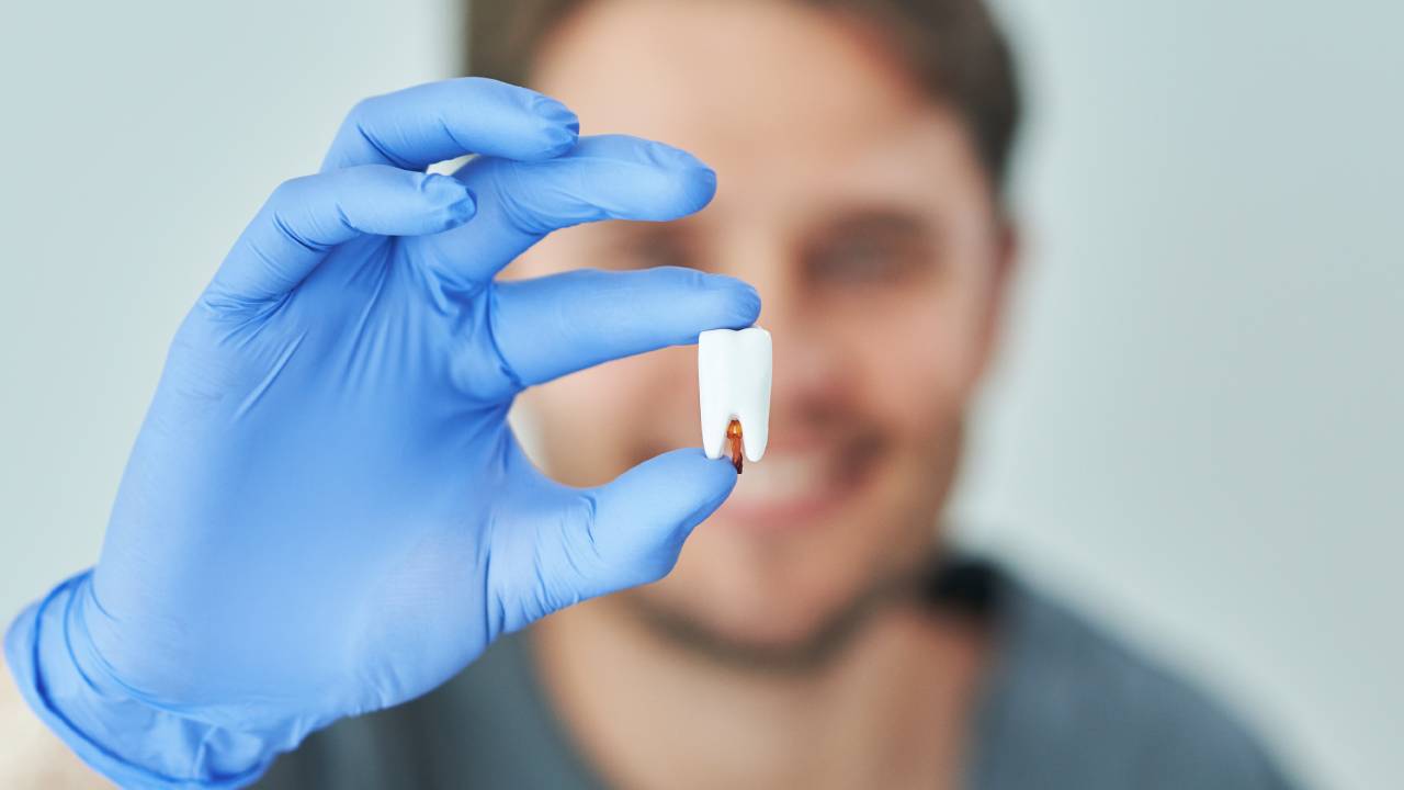
Understanding tooth damage
Before you schedule an evaluation for damaged teeth, it helps to know what structural issues your dentist will look for and why they matter. Tooth damage isn’t just about chips and cracks you can see. Underlying fractures, enamel wear, and weakened roots can compromise chewing stability and lead to more serious problems if left untreated. An evidence-based approach ensures your dentist recommends treatments that preserve function and durability rather than focusing solely on appearances [1].
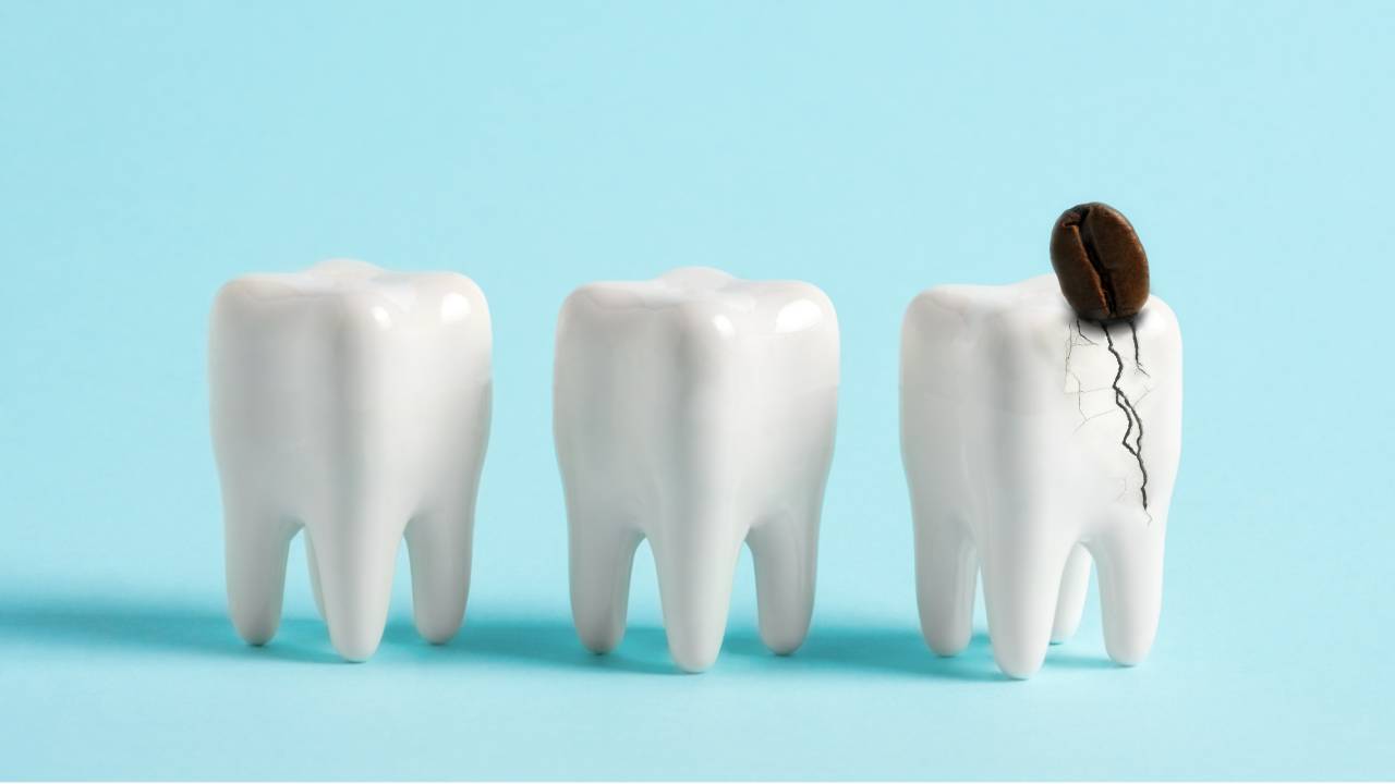
Damaged teeth fall into three broad categories: fractures, wear, and weakening.
Fractures range from superficial craze lines to deep vertical cracks extending below the gumline, allowing bacteria to invade the pulp chamber and potentially cause necrosis [2]. Wear patterns include attrition from grinding, erosion from acid exposure, and abrasion from aggressive brushing. Weakening often follows root canal therapy, large fillings, or chronic decay, leaving remaining tooth structure brittle and prone to failure.
Recognizing these types of damage early lets your dentist tailor an effective plan. Instead of waiting for pain or visible breaks, an in-depth assessment can catch hidden threats and guide preventive steps that maintain your natural dentition for years to come.
Recognizing damage symptoms
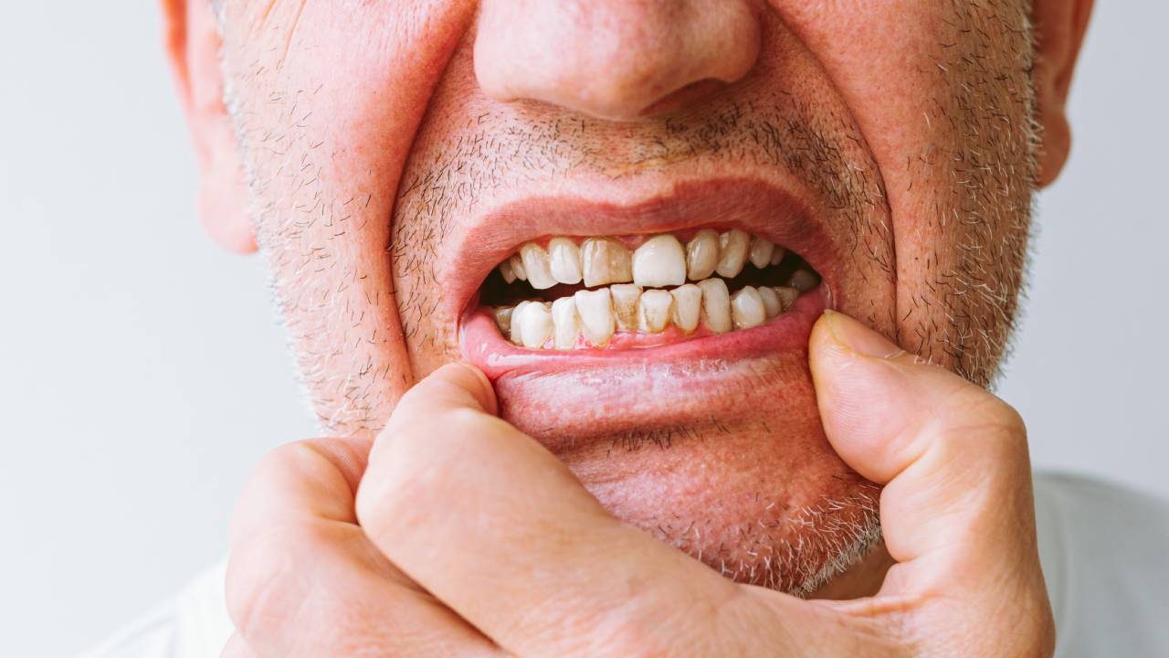
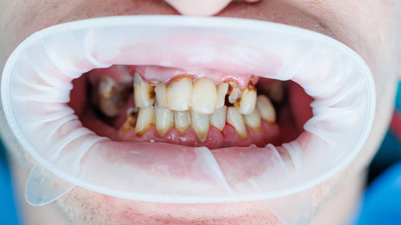
Pay attention to recurring signs that signal structural problems long before a tooth splits in two. Sharp sensitivity when you bite or release pressure is one of the most common red flags. You might feel a sudden jolt of pain while chewing on a seed or twinges when drinking something hot or cold. This often points to tiny cracks or enamel fractures that open under stress, then close and trap fluid, irritating nerve endings (biting pain cracked tooth dentist).
Visual clues are equally important. Look for small dark lines or shadows on the surface of the tooth, chipped edges or uneven wear facets, and areas where filling margins have separated from natural enamel. Even slight mobility—like a tooth that wobbles under gentle pressure—can indicate weakened roots or damaged supporting tissue.
When you notice these warning signs, it’s time to see a dentist for cracked enamel or a dentist for worn teeth. Early intervention can prevent a minor craze line from becoming a vertical root fracture requiring extraction.
Exploring diagnostic methods
Your dentist combines traditional exams with advanced imaging to form a complete picture of your tooth health. Understanding these methods helps you feel confident and informed before you arrive.
Visual and tactile exams
The first step is a close look under bright light, often enhanced with magnification or loupes. A mirror helps the dentist inspect all sides of each tooth, while a ball-ended explorer gently probes for rough spots or soft areas without risking enamel damage [3]. Periodontal probes measure gum pocket depth to detect subgingival cracks and early bone loss. Dentists also perform bite tests using tools like cotton rolls or tongue depressors to reproduce pain and isolate problem areas.
Radiographic imaging
X-rays remain a cornerstone of dental diagnostics, revealing hidden decay, root fractures, and bone abnormalities. Once your dentist reviews periapical and bitewing films, they may recommend a cone beam CT (CBCT) scan for complex cases. CBCT provides three-dimensional views of tooth roots and surrounding tissues, helping dentists locate cracks that elude standard X-rays. Although exposure is higher than 2D radiographs, the detailed information can be critical when planning a conservative restoration.
Emerging technologies
Newer non-invasive tools are transforming crack detection and enamel assessment. Optical coherence tomography (OCT) uses infrared light to reveal micro-cracks near the surface, while laser ultrasonic testing sends sound waves through enamel to detect structural flaws. Although these methods aren’t yet in every practice, they show promise for early diagnosis without exposing you to additional radiation [2].
| Method | Description | Strengths | Limitations |
|---|---|---|---|
| Periapical X-ray | Intraoral radiograph of individual teeth |
Low radiation, widely available | 2D view may miss fine cracks |
| Cone beam CT (CBCT) | 3D volumetric imaging | Detailed root and bone visualization | Higher cost and radiation dose |
| Optical coherence tomography | Infrared light scanning | Detects micron-level surface cracks | Limited penetration depth |
| MicroCT | High-resolution tomography | Research gold standard accuracy | Not practical for routine clinical use |
Preparing for your evaluation
Gathering a clear symptom history and medical background lets your dentist focus on the root causes of damage rather than symptoms alone. Bring a list of medications, note any history of trauma or grinding, and highlight previous restorative work such as large fillings or root canal therapy. Mention habits like nail-biting, ice chewing, or acid reflux, since all can contribute to enamel erosion and structural weakening.
Being honest about your daily routines and discomfort patterns allows your dentist to design treatment that strengthens your teeth against future wear and fractures.
If anxiety or gag reflex is a concern, let your practice know in advance. They may offer desensitization techniques or mild sedation so you can relax during both the examination and any necessary imaging.
Experiencing the assessment process
When you arrive, your dentist or hygienist will review your records and discuss your chief complaints. Expect questions about the onset and frequency of pain, triggers, and any changes you’ve noticed in tooth appearance. This interview often precedes the hands-on exam by a few minutes, ensuring everyone is aligned on your top priorities.
During the visual and tactile check, the dentist will use bite-and-release tests to isolate crack-related pain, percussion tests to assess periodontal health, and vitality tests (cold, heat, or electric pulp testing) to gauge nerve status. You may feel brief sensations of cold or tingling, which help map out areas of pulp irritation. If imaging has already been completed, your dentist will review the scans chairside, pointing out key findings that influence your repair strategy.
In some cases, your dentist will recommend a follow-up appointment just for 3D imaging or specialized diagnostics. This two-step approach prevents unexpected treatment decisions during your initial visit and gives you time to review financing or sedation options if needed.
Considering treatment options
After a thorough evaluation, your dentist will outline restorative pathways that balance durability, function, and esthetics. Your personal goals and budget both factor into whether you opt for a conservative approach or a more extensive solution.
Bonding with composite resin can seal minor cracks and rebuild worn enamel, preserving most of your natural tooth. For deeper fractures or weakened roots, onlays or full crowns distribute bite forces more evenly and protect the pulp from bacterial invasion. If a crack extends below the gumline or root strength is compromised, treatments like crown lengthening, root amputation, or even dental implant placement may become necessary [1].
Choosing a fixed restoration often yields better long-term performance and comfort than a removable appliance. Yet even with the strongest crown, your dentist will stress the importance of night guards if you grind, and targeted oral hygiene to prevent recurrent decay under margins.
Talk with a damaged tooth treatment dentist about the pros and cons of each option. You can also explore an assessment for tooth repair if you want a second opinion before committing to a specific procedure.
Maintaining long-term health
No restoration can outlast excellent preventive care. Once your damaged teeth are stabilized, commit to a routine that guards against future wear and fractures. Use a soft-bristle brush, low-abrasive toothpaste, and gentle circular strokes. Limit acidic snacks and beverages, and rinse your mouth after consuming citrus or soda. If grinding or clenching is part of your profile, wear a well-fitted occlusal guard nightly.
Regular check-ups (every six months or as recommended) let your dentist catch early signs of new damage and monitor existing restorations. Periodic bitewing X-rays ensure hidden decay doesn’t compromise bond margins, and digital scans can detect subtle shifts in wear patterns over time.
By partnering with a dentist for weakened teeth and staying proactive, you’ll safeguard your smile’s function and resilience for years to come.
When you understand what an evaluation for damaged teeth entails, you approach your appointment with confidence. You’ll know why each test matters, how to prepare, and what restorative options your dentist may recommend. That clarity empowers you to make informed decisions, preserve your natural dentition, and maintain comfortable, stable chewing long term.