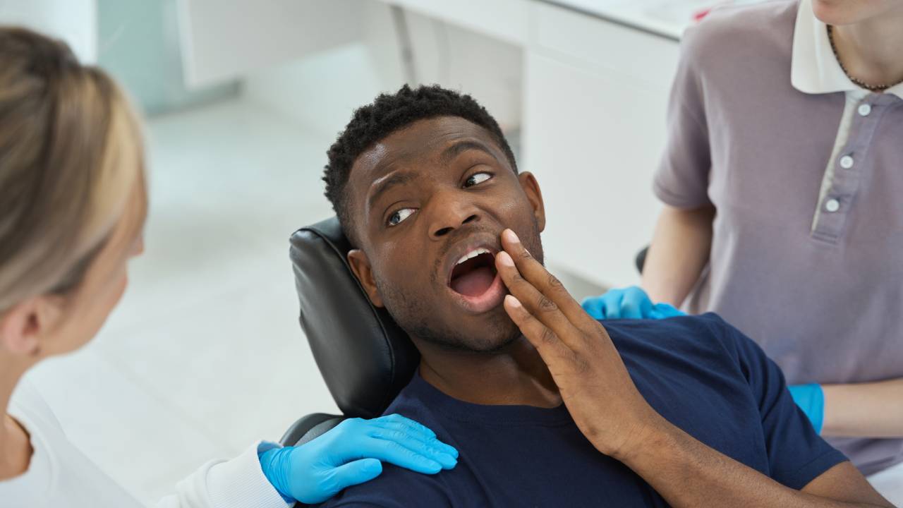
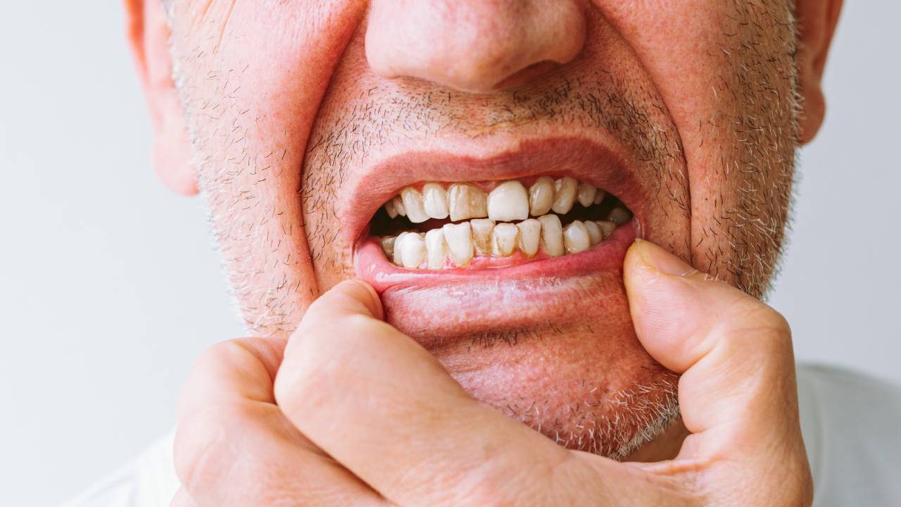
Missing a tooth can affect more than your smile. When you consult a missing tooth evaluation dentist, you start a process that safeguards your oral health and function. This guide walks you through every step of evaluation and planning so you can restore chewing stability and protect your bone.
By acting promptly, you avoid complications like bone resorption or shifting teeth. Bone loss begins within the first three to six months after extraction when your jaw stops receiving chewing pressure [1]. An early missing teeth dental evaluation gives you and your dentist a clear roadmap for tooth replacement planning.
Your evaluation sets the foundation for lasting restoration.
Why early evaluation matters
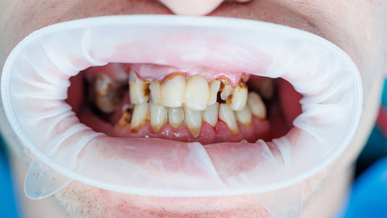
After tooth loss, the body immediately begins remodeling the jaw, which can lead to significant bone shrinkage if left unattended. In addition to the cosmetic impact, a narrowing ridge complicates the precise placement of an implant or a snug bridge fit. By securing a prompt assessment for missing teeth, you empower your dentist to preserve vital bone and simplify later restorative steps.
Consider the case of a patient who waited a year before replacing a molar. By the time she sought treatment, her ridge had atrophied to the point where a sinus lift and multiple grafts were needed before an implant could be placed. These additional surgeries extended her timeline by months and increased overall cost and discomfort.
In contrast, timely intervention—such as socket grafting immediately after extraction—can maintain ridge height and width. Studies show that placing graft material in the socket preserves up to 90 percent of original bone volume [1]. Acting within the first six months is your best defense against complex corrections later.
Preparing for your visit
Well before your appointment, gather relevant documents: recent X-rays, insurance details, and a list of current medications. If sedation dentistry or anxiety control is a consideration, mention this when you schedule. Many clinics offer guided tours or virtual consultations so you can familiarize yourself with the office layout and equipment ahead of time.
On the day of your visit, arrive a few minutes early to complete any remaining paperwork. Bring questions about timelines, cost breakdowns, and follow-up care. This preparation ensures that your dentist consultation for tooth loss moves efficiently and covers every concern you have.
Your medical history is equally important. Conditions like diabetes or osteoporosis can influence healing, just as certain blood thinners may require adjustments before grafting or implant surgery. Clear communication about your health background allows for a safe, personalized treatment plan.
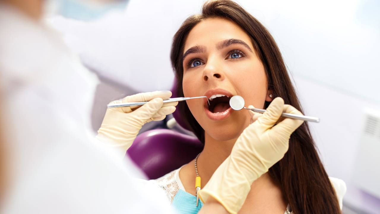
Overview of evaluation process
A systematic approach allows your dentist to assemble a detailed picture of your oral condition. The process generally unfolds in three phases: clinical examination, imaging diagnostics, and periodontal assessment. Each component contributes critical data for planning your replacement.
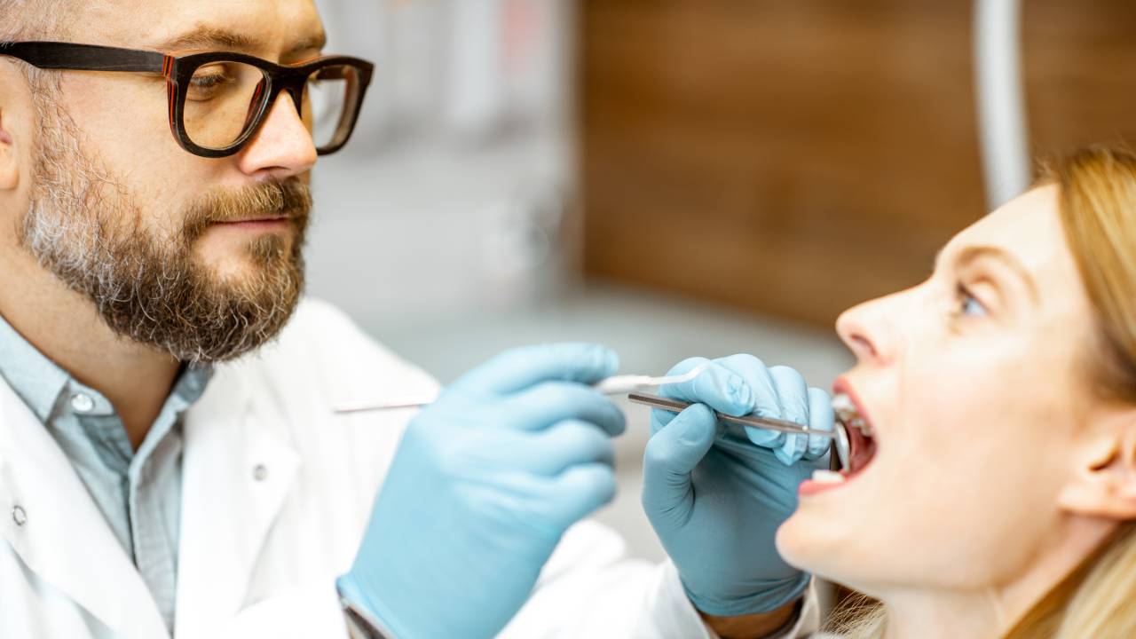
Clinical examination
Your dentist begins with a hands-on inspection of the extraction site and nearby teeth. They will measure the width of the ridge, assess any soft tissue irregularities, and check for signs of infection or lingering inflammation. Palpating the bone ridge helps determine if immediate grafting is required or if you have sufficient volume for an implant.
Imaging assessment
High-resolution digital X-rays, including periapical and panoramic images, provide a two-dimensional look at bone levels and adjacent structures. If your case demands unparalleled precision, a 3D CBCT scan maps bone density and vital anatomical features in three dimensions. These images guide the selection of implant length and diameter, helping avoid nerves and sinuses [2].
| Imaging type | Purpose | Benefits |
|---|---|---|
| Periapical X-ray | Detailed view of root and bone level | High resolution for diagnosing localized bone loss |
| Panoramic X-ray | Broad overview of the jaws and surrounding areas | Captures the full arch, useful for planning extractions or multiple tooth replacements |
| 3D CBCT scan | 3D volume assessment of bone density and shape | Precise measurement for implant positioning and identifying critical anatomical structures |
| Occlusal X-ray | Inspection of full dental arch | Evaluates developmental issues or fractures in the jaw arch [2] |
Periodontal charting
By measuring gum pocket depths around each tooth, your dentist identifies areas of active gum disease or bone loss. Pockets deeper than three millimeters may require scaling or deep cleaning before any restorative work. A healthy periodontal foundation not only supports the final restoration but also improves long-term stability.
Assessing bone health
Evaluating the quality and quantity of your jawbone is a critical determinant of which options you can pursue. Bone density is often classified into types D1 through D4, where D1 represents very dense bone more common in the front of the mouth and D4 indicates softer bone typically found in the posterior. This classification informs decisions on implant design and placement torque.
If your bone is insufficient, your dentist may recommend augmentation techniques. Socket preservation uses graft material immediately after extraction to prevent collapse. Ridge splitting gently expands a narrow ridge without adding donor material. In more complex cases, guided bone regeneration employs barrier membranes and synthetic or natural grafts to rebuild large defects [1].
Bone graft procedures can also draw on your own bone harvested from areas like the chin or ramus, or employ high-quality allografts. Deciding which approach suits you best depends on the size of the defect, your overall health, and your timeline for restoration.
Examining gum condition
The appearance and health of your gingiva are equally vital for a successful restoration, especially in the aesthetic zone. Thin or receding gum tissue may require a connective tissue graft to create a natural emergence profile around an implant crown. Without proper soft tissue volume, metal edges or abutments can become visible over time.
During your evaluation, the dentist checks for signs of inflammation, bleeding, and recession. They will assess tissue thickness and contour to predict how your gums will adapt around a permanent restoration. In some cases, a simple tissue punch or minor gum lift provides better symmetry and easier cleaning for long-term maintenance.
Ensuring healthy gums before moving forward reduces the risk of peri-implantitis or compromised bridge margins. By integrating gum health into your treatment plan, you secure both function and aesthetics in the years that follow.
Evaluating bite stability
Your bite, or occlusion, determines how forces from chewing distribute across teeth and restorations. A missing tooth can trigger tilting, drifting, or overeruption of opposing teeth in just a few months. Your evaluation will include an occlusal analysis, where bite impressions or digital scans reveal contact points and force distribution.
If you have experienced jaw pain, clicking, or muscle fatigue, your dentist may screen for temporomandibular disorders (TMD) during this phase. Addressing bite collapse early prevents undue stress on your new restoration and helps maintain joint health. Splints or night guards might be prescribed temporarily to stabilize your bite before final prosthetic work.
Correcting your occlusion often involves minor adjustments to existing teeth or even orthodontic realignment. A dentist for bite collapse from missing teeth collaborates with specialists to restore balanced function and protect both natural teeth and future restorations.
Exploring treatment options
After completing your comprehensive evaluation, your dentist will present a range of replacement pathways. Dental implants rank highest in longevity and bone preservation, as the titanium post stimulates the jaw similarly to a natural root [3]. Implants can support single crowns, bridges, or even snap-in dentures, offering versatility for one missing tooth or a full arch.
Fixed bridges rely on adjacent teeth for support, offering a shorter treatment timeline but requiring reduction of healthy tooth structure. Resin-retained bridges use a winged framework bonded to neighboring teeth, minimizing enamel removal but sacrificing some durability [4]. Removable partial or complete dentures remain cost-effective for multiple missing teeth or limited bone conditions [3].
Your dentist will walk you through advantages, drawbacks, and approximate costs, guiding you as a replacement options for missing teeth dentist. Budget considerations, healing time, and aesthetic goals all factor into the final decision.
Planning your replacement timeline
Understanding each stage in your journey helps you plan time off work and personal commitments. If you undergo socket grafting, anticipate a healing period of three to four months before implant placement. After implant insertion, osseointegration usually takes another three to six months, depending on bone quality and location.
For patients in the smile zone, many practices offer immediate provisional crowns to preserve aesthetics. These temporaries help shape gum tissue and maintain appearance while the site heals. You will return for a series of follow-up visits to adjust your provisional, capture final impressions, and verify bite before crafting the definitive crown or bridge.
Tracking your progress with a tooth replacement consultation dentist ensures that healing milestones are met. Should any delays occur, due to insufficient bone growth or soft tissue concerns, your dentist can adapt the timeline and explore interim solutions like removable flippers or partial dentures.
Understanding professional consultation
A successful outcome hinges on clear dialogue between you and your dental team. During your dentist consultation for tooth loss, express any fears, financial concerns, or aesthetic preferences. Ask for a written treatment plan with estimated dates, fees, and alternative paths if unexpected issues arise.
Many dentists employ digital smile design software to simulate post-treatment results, helping you visualize how your smile will appear. Obtaining a second opinion from a specialist, such as a periodontist or prosthodontist, can also add confidence in your chosen approach. Your collaborative treatment plan should align with both your functional needs and your vision for a restored smile.
Partnering with a dentist for tooth loss treatment ensures that your plan evolves alongside your healing milestones and long-term goals.
Maintaining oral health long term
Once your final restoration is in place, the work shifts to preservation. A rigorous home care routine combined with professional oversight reduces risks like peri-implantitis and recurrent decay. To keep your smile healthy, follow these steps:
- Brush twice daily with a soft-bristled brush and fluoride toothpaste, focusing around implant crowns and bridge abutments
- Floss every day using specialized floss or interdental brushes to clean beneath restorations
- Rinse with an antibacterial mouthwash to control harmful bacteria around grafted areas
- Attend routine cleanings and exam appointments every six months for radiographic monitoring of bone levels
In addition, maintain open communication with your dentist for long term tooth replacement or a functional tooth replacement dentist. Report any discomfort, looseness, or change in appearance immediately. Proactive care preserves both function and aesthetics, allowing you to enjoy a stable, confident smile for years to come.
By following this expert guide and partnering closely with your missing tooth evaluation dentist, you lay the groundwork for seamless tooth replacement planning. Early evaluation, thorough diagnostics, and tailored timelines come together to restore your smile, chewing ability, and long-term oral health.