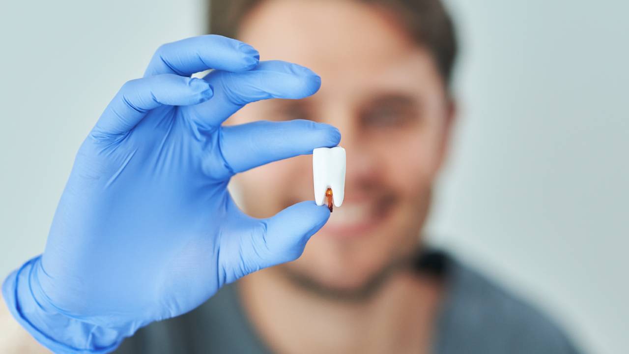
When you visit a tooth damage assessment dentist, you’ll find that their goal is to preserve your natural tooth structure by spotting fractures, wear, or weakening before they worsen. A thorough evaluation can mean the difference between a simple protective treatment and an invasive procedure like a root canal or extraction. In this guide, you’ll learn what your dentist looks for, how they assess different types of damage, and what you can do to protect your smile for the long term.
Structural tooth damage
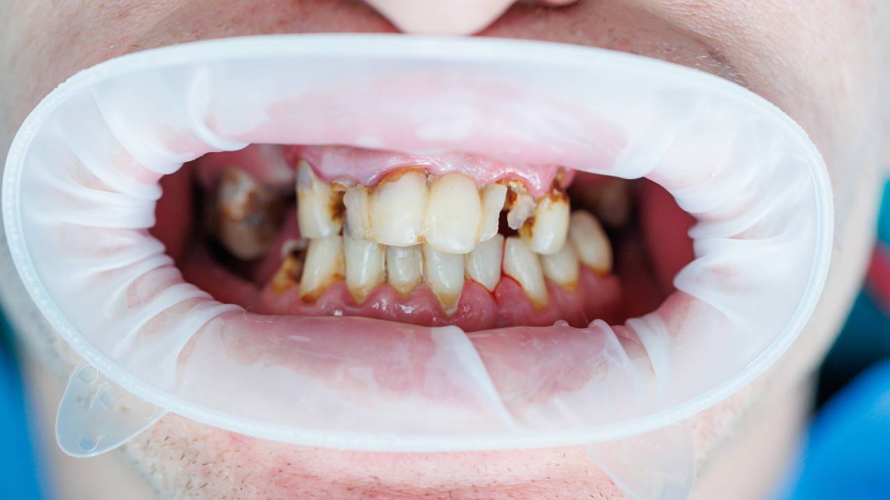
Teeth can suffer structural harm in several ways, each requiring a tailored approach to diagnosis and treatment. Understanding the differences helps you communicate symptoms clearly and set realistic expectations for repair.
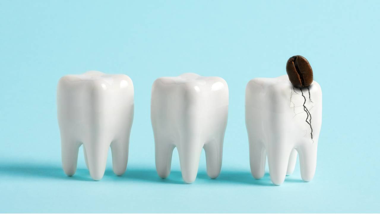
Fractures and cracks
Cracked enamel or deeper fractures interrupt the continuity of your tooth’s surface, creating a path for bacteria to invade. You might notice sharp pain when biting down or sensitivity to temperature changes. If you suspect a crack, scheduling an appointment with a dentist for cracked enamel or a dentist for tooth fracture ensures a focused evaluation, often including bite tests and magnified inspection.
Wear and erosion
Excessive wear can come from bruxism (teeth grinding), acidic diets, or abrasive brushing. Over time, enamel thins and dentin becomes exposed, leading to increased sensitivity and risk of decay. If your dentist spots flattened cusps, cupping on chewing surfaces, or glossy patches of eroded enamel, they may recommend night guards or dietary changes to slow progression and protect exposed areas.
Weakening and demineralization
Early mineral loss, often invisible to the naked eye, can compromise tooth strength long before a cavity forms. Your dentist may detect these weakened zones through careful tactile probing or specialized fluorescence tools. Addressing demineralization with fluoride varnishes or sealants often halts progression and restores resilience without drilling.
Importance of early assessment
Catching structural issues early preserves more of your natural tooth and reduces the need for complex treatments. Dental exams are critical for detecting decay before it damages deeper layers of your tooth, helping you avoid invasive procedures like root canals or extractions [1].
Early assessment also uncovers hidden gum disease, which can undermine the support around a structurally compromised tooth, and even screens for oral cancer—conditions you may not notice until they become severe [1]. By prioritizing evaluation, you preserve both function and aesthetics over the long term.
Evaluation methods dentists use
Dentists combine clinical skills with targeted tests to pinpoint the exact nature of your tooth damage.
Visual and tactile examination forms the foundation of every assessment. Using a mouth mirror and explorer, your dentist scans each surface for discoloration, pits, or rough edges that signal underlying issues [2].
Functional tests help reveal non-obvious fractures. You might be asked to bite gently on a specialized tool or perform lateral movements; pain or “clicks” during these maneuvers can indicate cracks extending into the dentin or root.
Thermal and percussion tests further clarify pulp vitality and detect microfractures. A cold stimulus applied to suspect teeth gauges nerve response, while light tapping identifies areas of sensitivity that could suggest structural compromise or inflammation.
Diagnostic tools and technologies
Advanced imaging and detection devices enhance accuracy, often revealing damage that escapes a standard exam.
Digital X-rays
Digital radiography delivers low-radiation images with high resolution, uncovering hidden cavities, cracks, and bone loss. Your dentist can spot decay between teeth or beneath existing restorations long before you feel any discomfort [3].
Intraoral cameras
These small, high-definition cameras provide real-time visuals of enamel erosion, craze lines, and early decay. You’ll see the same images your dentist views, so you understand exactly where and how your tooth is compromised.
Laser fluorescence
By measuring changes in tooth density, laser fluorescence devices detect demineralized areas invisible on X-rays or to the naked eye. This technology is especially helpful for diagnosing early-stage decay and monitoring repair efforts without invasive probing.
Classification systems overview
Standardized frameworks help your dentist quantify lesion severity and plan appropriate interventions.
| System | Focus | Key features | Limitations |
|---|---|---|---|
| ICDAS II | Caries staging from early to cavitation | 0–6 scoring, includes lesion activity and location | Requires training and detailed charting [4] |
| ADA Caries Classification System | Lesion categories by anatomy and activity | Sound, initial, moderate, advanced categories | Less granular staging than ICDAS [4] |
| G.V. Black classification | Historical cavity preparation design | Six classes based on cavity location | Does not detect early lesions, may lead to over-preparation [4] |
| Decayed-Missing-Filled (DMF) index | Epidemiologic caries experience | Counts decayed, missing, and filled surfaces | Cannot distinguish active vs. inactive lesions [4] |
Each system guides decisions—from whether to apply a sealant [5] to how aggressive a restoration should be.
Preparing for your dental exam
Before your visit, compile a concise history of your symptoms—when you first noticed sensitivity or discomfort, and any recent trauma.
Bring any previous X-rays or records, especially if you’ve seen another provider for a dentist for tooth trauma evaluation or fractured tooth dentist appointment. Clear communication helps streamline your assessment and ensures your dentist has the full picture.
Preventing further deterioration
Halting progression of cracks, erosion, or demineralization hinges on consistent home care and lifestyle adjustments. Maintain twice-daily brushing with fluoride toothpaste, floss daily to remove interdental plaque, and limit acidic or highly abrasive foods. Custom night guards protect against bruxism, while topical fluoride or sealants strengthen vulnerable surfaces. By following these recommendations, you’ll extend the lifespan of your natural teeth and reduce the need for extensive repairs.
Planning treatment options
After a comprehensive assessment, your dentist will outline treatment paths tailored to the type and severity of damage. Minor enamel cracks may only require smoothing and monitoring, while deeper fractures often call for bonded restorations, onlays, or full crowns. Weakened teeth with significant decay might benefit from root canal therapy followed by a protective crown to restore both function and strength.
Discuss concerns such as cost, durability, and appearance—your dentist can help you weigh options like composite bonding versus porcelain inlays. For personalized guidance, consider an assessment for tooth repair or consult a tooth structure damage dentist who specializes in restorative solutions.
By understanding how your tooth damage assessment dentist evaluates structural harm—and by taking proactive steps to protect enamel and dentin—you empower yourself to make informed decisions and safeguard your smile for years to come.