Expertise provided by Dr. Moshos, Owner
You’ve been told you need a cone-beam CT scan before your dentist can plan an implant, finish a root canal, or figure out what’s going on with that molar in the back, and the first thing you wonder is whether this is really necessary or just an expensive upsell. That’s a fair question, so here’s a straight answer. At River’s Edge Dental in Mooresville, NC, every new adult patient receives a cone beam CT dental imaging scan, and it isn’t there to pad a bill. It’s there because flat X-rays can leave out information that may help decide whether a treatment might be appropriate for your situation.
This guide walks you through what the scan is, when it may change your treatment plan, how much radiation you receive, what you might pay, and what the appointment feels like if you’re already nervous about the dentist. If you’re anxious, you are not alone, it’s very common, and nothing about this scan requires you to be brave.
What Is Cone Beam CT Dental Imaging, and How Is It Different From Your Regular X-Rays?
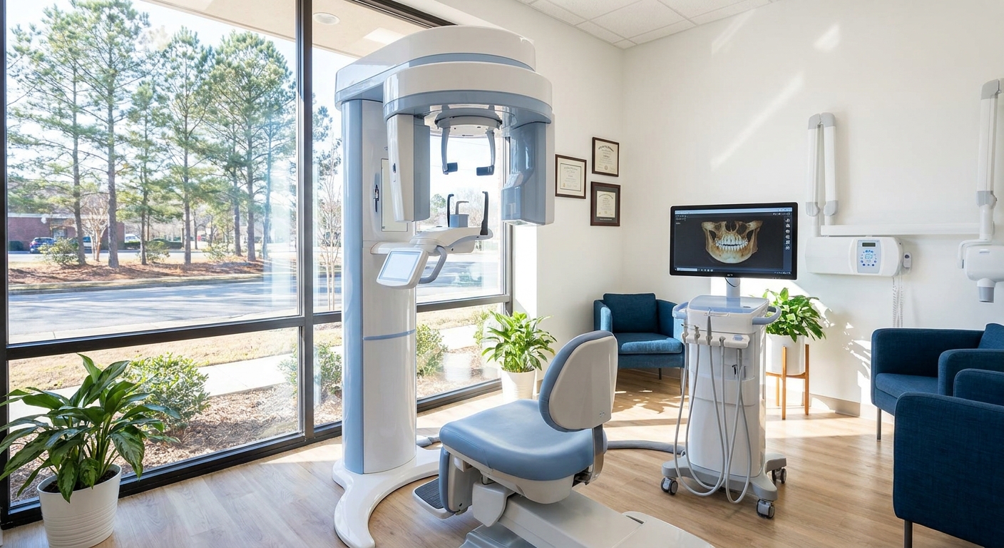
Cone beam CT, short for cone beam computed tomography, is a 3D X-ray of your teeth, jaws, nerves, and the bone around them. A regular dental X-ray gives you one flat picture, but a cone-beam machine circles your head once and builds a full 3D model that your dentist can turn, slice, and measure on a screen.
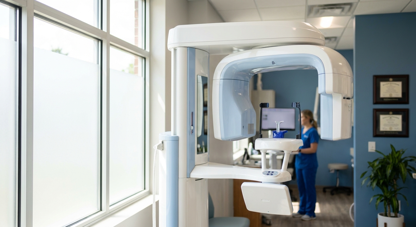
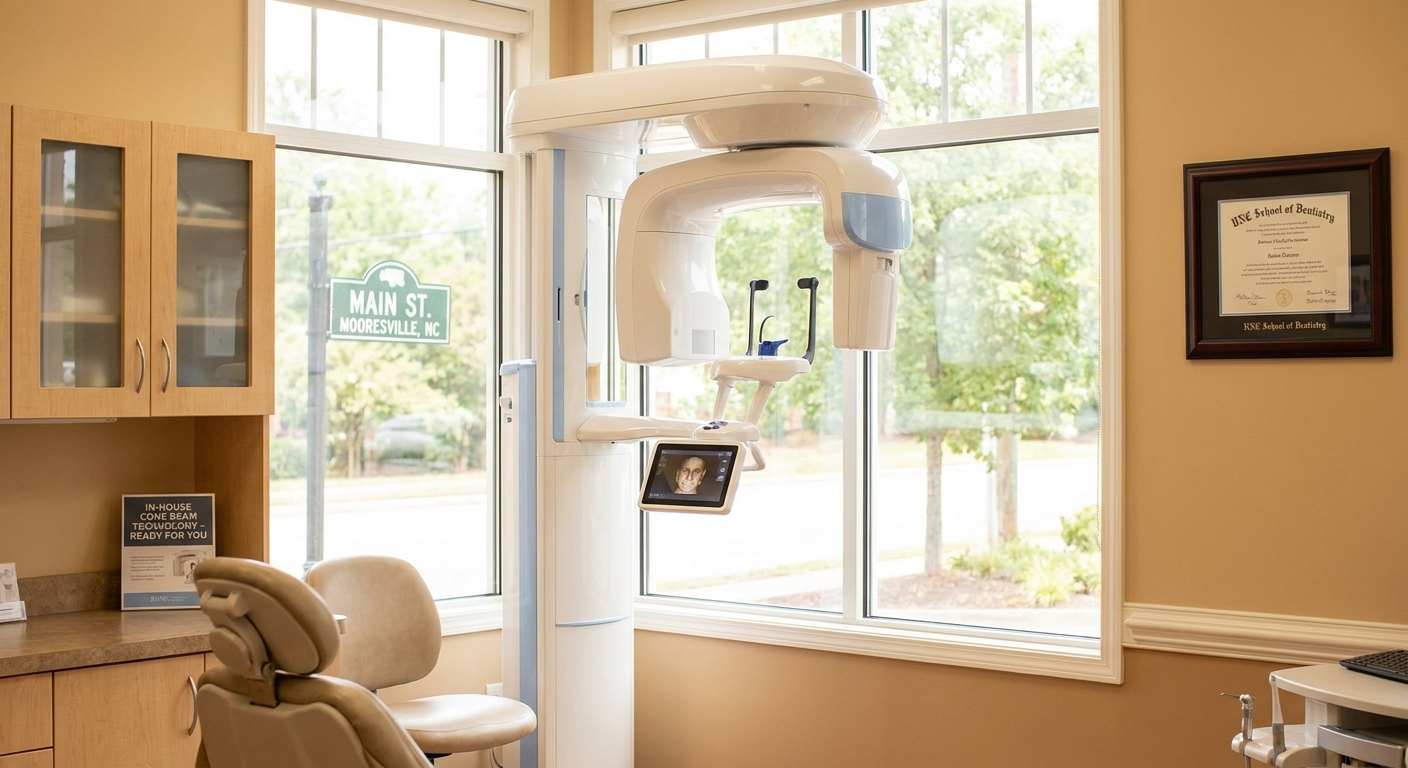
The name describes how the machine works. A regular medical CT scanner uses a thin, fan-shaped beam and takes many passes, while a dental cone-beam unit uses a cone-shaped beam that captures the whole area in a single rotation around the head. You stand or sit still while the arm of the machine moves around you, and because River’s Edge Dental has the unit in-house, the dentist can pull up the 3D image on a monitor in the same room.
Here’s a simple way to picture the difference. A traditional 2D X-ray is like a photo of a loaf of bread taken from the top, so you can see the shape but you can’t see what’s inside each slice. A cone-beam scan lets you cut the loaf and look at every piece from any angle. On a flat X-ray, everything stacks on top of everything else, so one root can hide behind another, the sinus can overlap your upper molars, and bone that looks solid from the front can turn out to be thin from the side.
None of this means your regular X-rays go away. Bitewings are still commonly used to check for cavities between teeth, evaluate the margins on a crown or filling, and monitor results after treatment. The 3D scan doesn’t replace any of that. It can help answer questions a flat picture may not address, mostly about depth, width, and what’s hiding behind something else.
Why Does River’s Edge Dental Scan Every New Adult Patient in 3D?
River’s Edge Dental scans every new adult patient in 3D because findings that may affect treatment decisions are sometimes not visible on 2D imaging. The three that come up frequently include root complications, additional canals, and the amount of bone available for an implant.
When a new adult patient sits down in the chair, they bring years of dental history nobody at the practice saw happen: an old crown, a root canal from a dentist two moves ago, or a tooth that’s been “a little off” for a while. A 2D image might make all of that look fine, and then the 3D scan may show a root that appears compromised, a canal that wasn’t treated, or bone that has quietly thinned out. That information can help inform diagnosis, including whether a tooth might be saved, whether an implant has enough bone to work with, or whether a different approach may be worth considering.
It’s worth being upfront about something, too. Professional guidance from national dental and dental radiology organizations says imaging should be selected for each individual patient, not ordered out of habit. That’s a fair standard to hold any office to, including this one. The reasoning at River’s Edge Dental is that one baseline cone beam CT dental imaging scan at the start can provide more comprehensive information before a treatment plan is built, which may help reduce surprises that sometimes appear midway through treatment. If you have questions about why the scan makes sense for you, just ask. The dentist will walk you through it, and nobody is going to rush you.
This may be particularly relevant if you’ve had difficult experiences before. Patients who come in with dental anxiety often describe how uncomfortable it felt to be surprised in the middle of a procedure. A 3D scan doesn’t remove every surprise, but it may help reduce some of them. And the scan isn’t a tool for upselling. If there are a few craze lines on your front teeth and they’re not bothering you, that’s not a reason to cut those teeth for crowns. The scan can show what’s there, so the plan you agree to reflects what the dentist actually observes.
When Does a 3D Scan Actually Change Your Treatment Plan?
A 3D scan may inform the plan most often in three situations: when you’re thinking about replacing a missing tooth with an implant, when you’re trying to figure out why a tooth with an old root canal still bothers you, and when there’s hidden bone loss to measure. In each case, the scan provides a measurement or a finding that a flat X-ray may not clearly show.
Clinical reviews describe these same core uses for dental cone beam computed tomography (CBCT), including implant planning, evaluating root canal anatomy, and assessing bone. Bone loss around teeth deserves its own mention. On a 2D image, you only see bone levels from the front and back, but a cone-beam scan shows the cheek side and the tongue side too, and that’s often where bone loss hides. When it shows up there, it can affect whether a tooth may have a good long-term outlook, and it’s something you might want to know before you invest in a crown on that tooth.
Here’s how the scan may inform the two decisions patients ask about most: implant planning and root canal problems.
Implant Planning: Measuring Bone Height, Width, and Nerve Position
Because the scanner is in-house, River’s Edge Dental plans single-tooth implants right in the office instead of sending you elsewhere for imaging. In some cases where bone appears sufficient, a same-day implant may be possible, though most implant appointments are scheduled as a separate visit so you aren’t rushed.
The scan can be particularly helpful because an implant needs to fit the bone you actually have, not the bone a flat X-ray suggests. The practice at Rivers Edge Dental works from specific measurements. The narrowest implants are 3.5mm wide, and placing one typically calls for at least 6mm of bone width, with 7mm often preferred, so healthy bone can surround the implant on both sides. The shortest implants are 8.5mm long, and they generally need at least 10.5mm of bone before reaching a vital structure like the alveolar nerve that runs through your lower jaw. A flat X-ray can’t reliably show bone width at all, and it can blur where that nerve sits. The 3D scan can show both, which may help in selecting implant size with a clearer margin of safety.
Upper back teeth bring a different challenge, which is the sinus. Above your upper molars sits an air space, and sometimes the floor of that sinus sits low, leaving less bone height than an implant might need. When the scan shows limited height, River’s Edge Dental can perform a small sinus lift using specialized reverse-cutting Versah burs, which shape and compact the bone to gain height in some patients. You’ll know up front if that step may be part of your plan instead of finding out later.
Either way, you go into the implant conversation with real measurements, not guesses. You’ll have time to think it over, review costs, and come back calm before anything moves forward, and the scan is what helps make that kind of honest, unhurried conversation possible.
Root Canal Cases: Finding Extra Canals and Compromised Roots
A cone-beam scan may help explain why a tooth that “looks fine” on a flat X-ray still hurts. The two most common reasons include a canal that wasn’t found and treated, and a compromised root that a 2D image can’t separate from the structures around it.
Picture a tooth that had a root canal years ago. It’s still tender when you chew, and your last X-ray didn’t show anything. Some molars have an extra canal that hides directly behind another one on a flat image, and if that canal was missed, bacteria can stay inside the tooth regardless of how good the rest of the work looked. On a 3D scan, the dentist can scroll through the root slice by slice and may see that missed canal, or see a dark area of bone at the tip that suggests the area never fully healed.
That finding can inform a real decision. Some teeth may be candidates to have the root canal redone, and in more complex cases you may be referred to a local specialist. Other teeth appear to have a fractured or compromised root that may not respond predictably to retreatment, and for those it may make more sense to consider removing the tooth and discussing replacement options. It can be helpful to know that before you spend money on a new crown for a tooth that may not last.
How Much Radiation Does a Cone-Beam Scan Really Give You?
If you’re wondering about radiation, that’s a good question to ask. The radiation exposure from a dental cone-beam scan is higher than a standard set of dental X-rays but generally lower than a conventional medical CT. The exact dose depends on how large an area is scanned and how the machine is set, and a small, focused scan sits at the low end.
Federal regulators explain that dental cone beam computed tomography doses are higher than conventional dental X-rays and that the dose varies widely between machines and scan sizes. That’s why the size of the area scanned, called the field of view, matters so much. Scanning one section of the jaw uses less radiation than scanning both jaws and the sinuses, so a thoughtful approach involves selecting the smallest field of view that still answers the question.
That’s also the thinking behind current clinical recommendations on radiation safety in dentistry, which stress two ideas: every image should be justified, and every dose should be kept as low as practical while still giving a usable result. At River’s Edge Dental, the goal for a new adult is one clear baseline image, not a scan every visit, and a good 3D image can answer a question that would otherwise mean several more flat films from different angles and still a fair amount of uncertainty.
If radiation is part of why you’re nervous, say so, and the dentist can explain which area is being scanned and why. If you are or might be pregnant, let the Rivers Edge Dental team know before any imaging so you can talk about timing. And if you’re booking for the whole family, the routine 3D scan is only for new adult patients, so your kids won’t be lined up for one just because everyone was scheduled together.
What Will the Scan Cost, and What Happens at the Appointment?
Cost is a common worry. Many dental plans don’t pay for a 3D scan as a routine benefit, so expect some out-of-pocket cost. A dental cone-beam CT scan generally runs from about $200 to several hundred dollars, depending on how much of the mouth and jaw is scanned, and the front desk at River’s Edge Dental can give you the exact fee when you call.
Coverage varies from plan to plan. Some plans treat cone-beam imaging as advanced diagnostic imaging and leave it to you, while others may help in specific situations, such as implant planning. River’s Edge Dental accepts UnitedHealthcare, Delta Dental, Aetna, Principal, and BlueCross BlueShield of North Carolina, and the team can check what your specific plan says about imaging before you come in. If the scan leads to treatment, you can talk through how to phase it so you get the most from your benefits, with your out-of-pocket share made clear at each step.
Here’s what the visit actually feels like. You check in and let the team know if you’re anxious, and everything moves at your pace. You’ll take off earrings, glasses, and anything removable in your mouth, since metal blurs the image. Then you stand or sit at the machine and bite gently on a small guide to keep your head steady. You won’t be inside a tube like a medical CT or MRI, because the arm simply moves around the outside of your head. There are no needles and no discomfort, and it feels a lot like a panoramic X-ray.
Then comes the part patients tend to appreciate most. Because the scan is taken in-house at Rivers Edge Dental, the dentist can pull up your 3D images during the same appointment and show you exactly what they see: your bone, your roots, and any areas of concern, along with why a particular treatment may be recommended and what your options are. If you need time, a written plan, or a phased approach, that’s fine. Walking out actually understanding your own mouth is the whole point.
Why Mooresville Patients Trust River’s Edge Dental With What Their Scan Shows
A scan is only as useful as the person reading it. At River’s Edge Dental, in-house cone-beam imaging comes with advanced implant training and a comfort-focused, anxiety-free approach, so you may receive clearer information without being handed off to someone else.
The dentist at River’s Edge Dental completed the MaxiCourse in implant dentistry at Augusta University about eight years ago. That training provides clinical background for reading cone-beam scans: evaluating whether bone width meets the 6mm to 7mm typically considered for placing even the narrowest implants, confirming clearance to the nerve, and noting when a low sinus floor or a compromised root might affect the plan. Having the imaging in-house also means you don’t need a second appointment at a separate imaging center, and you don’t have to wait for results to travel between offices.
River’s Edge Dental is an independent practice, not part of a large corporate group, and that shapes how technology gets used. The scan is there to support personalized care and a genuine connection with you, not to hit a quota. The Rivers Edge Dental practice is currently in the top three for a NORMY award, which recognizes dentists in Mooresville, and it has received Best Practice awards from Google based on its patient reviews. The practice didn’t nominate itself for those Google awards. They came from what patients wrote.
Patients come to Rivers Edge Dental from Mooresville and nearby Davidson, Cornelius, Kannapolis, and the Lake Norman area. Many of them put off care for years because of how they were treated somewhere else. If that’s you, the judgment-free part isn’t a slogan. It’s how the appointment actually runs, scan included.
What Else Do Patients Ask Before a Cone-Beam Scan?
Most questions come down to time, cost, comfort, and whether you truly need the scan. Here are short, direct answers to the ones that come up most in Mooresville.
How long does a dental cone-beam CT scan take?
The scan itself takes only seconds while you hold still. At River’s Edge Dental, the machine is in the office, so the dentist can review the images with you during the same appointment.
Is cone-beam CT covered by dental insurance?
Often it isn’t, because many plans classify it as advanced diagnostic imaging. Coverage depends on your plan, so the Rivers Edge Dental team can check your benefits ahead of time and tell you what to expect out of pocket.
How much radiation does a cone-beam CT scan deliver?
The radiation exposure is more than a standard set of dental X-rays and generally less than a conventional medical CT. The dose depends on how large an area is scanned, and the smallest field of view that answers the clinical question is typically the one that should be used.
What can dentists see on a cone-beam CT that they can’t see on regular X-rays?
A 3D scan can show compromised roots, additional canals, bone height and width measurements for implants, the position of nerves and the sinus, and bone loss on the cheek and tongue sides of teeth. Flat X-rays may not reveal these clearly.
Do I really need a cone-beam CT scan, or is it just an upsell?
River’s Edge Dental scans new adult patients because the scan may reveal concerns that could affect whether a treatment is appropriate and how it might proceed. If you’re unsure, ask the dentist to explain what they’re looking for in your case.
What does a cone-beam CT scan feel like?
It’s painless and non-invasive. You stand or sit still with your teeth resting on a small guide while the machine circles your head, and you won’t be enclosed in a tube.
If you’ve been putting off a tooth that just doesn’t feel right, or you’re weighing a bigger treatment and want honest numbers before you commit, schedule a new patient exam at River’s Edge Dental in Mooresville. Your comprehensive evaluation includes cone beam CT dental imaging, so you may see more clearly what’s happening beneath the surface before you agree to any treatment plan. One tip when you call: if a specific tooth has been bothering you, especially one with an old root canal or crown, tell the team which one when you book. That way the dentist knows where to look first on your scan, and you may leave your first visit with a clearer picture of that tooth instead of another “let’s wait and see.”
See What Traditional X-Rays Can’t Capture
If you’ve been told you need a complex procedure or simply want a more complete picture of your oral health, Cone-Beam CT imaging can provide the clarity you and your dentist need to move forward with confidence. Our team at Rivers Edge Dental in Mooresville uses this advanced 3D technology to plan treatment more precisely and help you understand exactly what’s happening beneath the surface. We’re here to answer your questions and discuss whether this imaging is right for your situation.
Every smile is different, which is why our personalized care begins with a thorough exam and a genuine connection. Your dentist will talk through your dental anatomy, health history, and goals so your comfort-focused treatment plan fits you.